Posterior Anatomy Of Rib Cage : Scielo Brasil Anatomical Variations And Congenital Anomalies Of The Ribs Revisited By Multidetector Computed Tomography Anatomical Variations And Congenital Anomalies Of The Ribs Revisited By Multidetector Computed Tomography : It depresses the lower rib cage.
Posterior Anatomy Of Rib Cage : Scielo Brasil Anatomical Variations And Congenital Anomalies Of The Ribs Revisited By Multidetector Computed Tomography Anatomical Variations And Congenital Anomalies Of The Ribs Revisited By Multidetector Computed Tomography : It depresses the lower rib cage.. Superiorly by the 12th rib and diaphragm. Lateral view of a pair of ribs articulating with the thoracic vertebrae. The ribs are a set of twelve paired bones which. Connective tissue of rib cage. Rib cage posterior view in humans, the rib cage, also known as the thoracic cage, is a bony and cartilaginous structure which surrounds the thoracic cavity and supports the pectoral girdle (shoulder girdle), forming a core portion of the human skeleton.
The posterior end of a typical rib is called the head of the rib (see chapter 7.3 figure 7.3.8). Check spelling or type a new query. The thoracic spine is unique from the cervical and lumbar spine because of the size and extent of the region and the articulations with the rib cage. Rib cage pain may be sharp, dull, or achy and felt at or below the chest or above the navel on either side. The 10th and 11th intercostal spaces are supplied by the posterior.
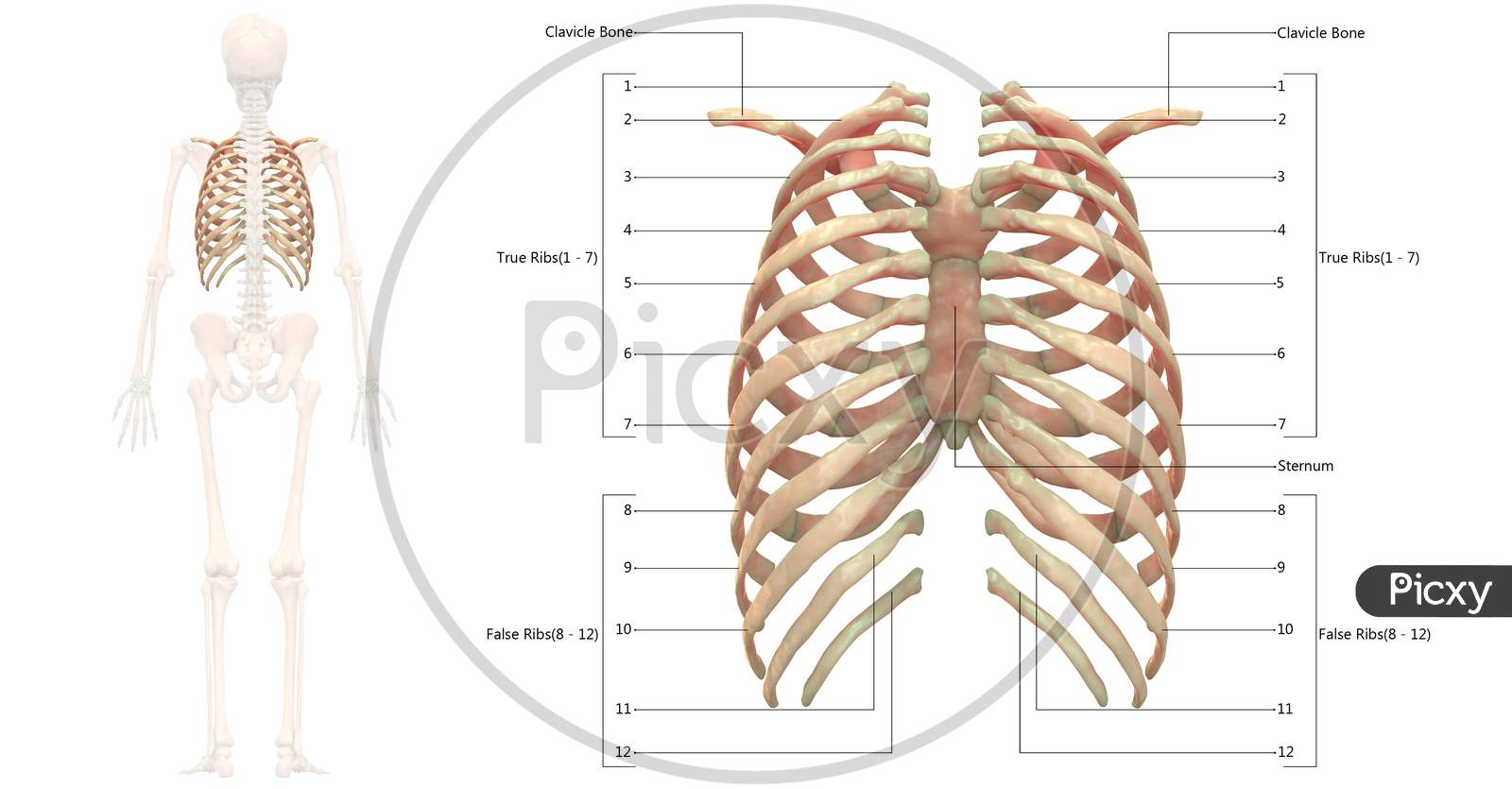
Chest Abdomen Pelvis Perineum Groin Trunk Back Thorax from slidetodoc.com It may occur after an obvious injury or without explanation. Ten of the twelve ribs connect to strips of hyaline cartilage on the anterior side of the body. Superiorly by the 12th rib and diaphragm. The thoracic cage (rib cage) is the skeletal framework of the thoracic wall, which encloses the thoracic cavity. The rib cage surrounds the lungs and the heart, serving as an important means of bony protection for these vital organs.in total, the rib cage consists of the 12 thoracic vertebrae and the 24 ribs, in addition to the sternum. Check spelling or type a new query. The anatomical terms of location are vital to understanding and using anatomy. Each of the upper 9 intercostal spaces is supplied by the three vessels;
This region articulates primarily with the costal facet located on the body of the same numbered thoracic vertebra and to a lesser degree, with the costal facet located on the body of the next higher vertebra.
Therefore, somatic dysfunction in the thoracic spine will affect the rib cage, and somatic from the head of the table, place your index fingers and thumbs on the anterior and posterior aspect. Posterior and inferiordirection and the posterior rib angle in an anterior and superior direction, decreasing the anteroposterior diameter of the rib cage. The angles of the ribs form the most posterior extent of the thoracic cage. It is innervated by the first four lumbar nerves, plus the twelfth thoracic nerve. Superiorly by the 12th rib and diaphragm. The 10th and 11th intercostal spaces are supplied by the posterior. A single posterior intercostal artery and the two anterior intercostal arteries. Doctors from medicinenet say that the middle and upper part of your spine contains 12 vertebrae that are attached to your rib cage. The posterior end of a typical rib is called the head of the rib (see chapter 7.3 figure 7.3.8). Connective tissue of rib cage. The ribs are a set of twelve paired bones which form the protective 'cage' of the. Rib cage anatomy the rib cage, shaped in a mild cone shape and more flexible than most bone sets, is made up of varying elements such as the thoracic vertebra, 12 equally paired ribs, costal cartilage, and held together anteriorly by the sternum. This region articulates primarily with the costal facet located on the body of the same numbered thoracic vertebra and to a lesser degree, with the costal facet located on the body of the next higher vertebra.
The tubercle, a bony protuberance on the posterior surface of the rib, articulates with the transverse process of the vertebra, forming the costotransverse joint ( fig. It depresses the lower rib cage. Gross anatomy there are 12 pairs of ribs which are separated by intercostal spaces. Check spelling or type a new query. Inserts at the xiphisternum and the 5 th to 7 th costal cartilages;
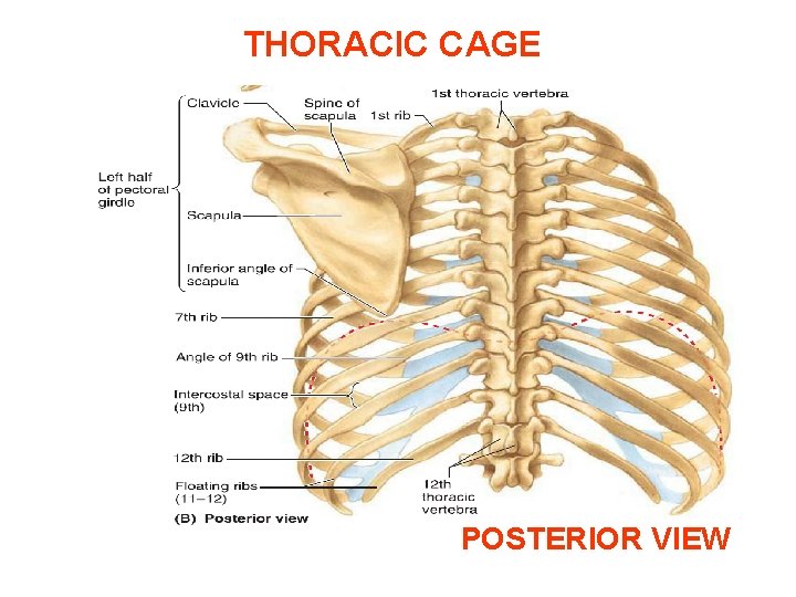
Image Of Human Skeleton System Rib Cage Bone Joints Described With Labels Anatomy Posterior View Xh791314 Picxy from images.picxy.com In the anatomical position, the angles align with the medial border of the scapula. Ninja nerds!join us in this video where we show the sternum and rib articulation anatomy through the use of a model. This is likened to the movement of a pump handle. Lateral view of a pair of ribs articulating with the thoracic vertebrae. It is innervated by the first four lumbar nerves, plus the twelfth thoracic nerve. This region articulates primarily with the costal facet located on the body of the same numbered thoracic vertebra and to a lesser degree, with the costal facet located on the body of the next higher vertebra. The rib cage surrounds the lungs and the heart, serving as an important means of bony protection for these vital organs.in total, the rib cage consists of the 12 thoracic vertebrae and the 24 ribs, in addition to the sternum. Just lateral to the tubercle is the angle of the rib, the point at which the rib has its greatest degree of curvature.
Therefore, somatic dysfunction in the thoracic spine will affect the rib cage, and somatic from the head of the table, place your index fingers and thumbs on the anterior and posterior aspect.
The ribs are a set of twelve paired bones which. The remainder of the rib is the body of the rib (shaft). At the chest, many rib bones connect to the sternum via costal cartilage,. The angles of the ribs form the most posterior limit of the thoracic cage and are in line with the medial border of the scapula. Each rib forms two joints: With each succeeding rib, from the first, or uppermost, the curvature of the rib cage becomes more open. Inserts at the xiphisternum and the 5 th to 7 th costal cartilages; The thoracic spine is unique from the cervical and lumbar spine because of the size and extent of the region and the articulations with the rib cage. Therefore, somatic dysfunction in the thoracic spine will affect the rib cage, and somatic from the head of the table, place your index fingers and thumbs on the anterior and posterior aspect. The ribs are a set of twelve paired bones which form the protective 'cage' of the. Check spelling or type a new query. The thoracic cage consists of the 12 thoracic vertebrae, the associated intervertebral discs, 12 pairs of ribs with their costal cartilages, and the sternum. Each of the upper 9 intercostal spaces is supplied by the three vessels;
With each succeeding rib, from the first, or uppermost, the curvature of the rib cage becomes more open. The 10th and 11th intercostal spaces are supplied by the posterior. Each of the upper 9 intercostal spaces is supplied by the three vessels; It is innervated by the first four lumbar nerves, plus the twelfth thoracic nerve. Ten of the twelve ribs connect to strips of hyaline cartilage on the anterior side of the body.
Thorax Bones Of The Rib Cage from www.anatomystandard.com Doctors from medicinenet say that the middle and upper part of your spine contains 12 vertebrae that are attached to your rib cage. The remainder of the rib is the body of the rib (shaft). The flexible (hyaline) cartilage, makes the breathing process easier. Lateral view of a pair of ribs articulating with the thoracic vertebrae. The tubercle, a bony protuberance on the posterior surface of the rib, articulates with the transverse process of the vertebra, forming the costotransverse joint ( fig. Diagram of human body, liver rib cage, rib cage diagram labeled, rib cage diagram numbered, rib cage diaphragm, rib cage heart, rib cage organs anatomy, rib cage pain, stomach, diagram of human body, liver rib cage, rib cage diagram labeled, rib cage diagram numbered, rib cage diaphragm, rib cage. The anterior and posterior intercostal arteries travel within the intercostal spaces, bordered by the lower edge of the rib above and the upper border of the rib below. The 10th and 11th intercostal spaces are supplied by the posterior.
The thoracic spine is unique from the cervical and lumbar spine because of the size and extent of the region and the articulations with the rib cage.
This muscle assists in depression of the ribs. The flexible (hyaline) cartilage, makes the breathing process easier. Just lateral to the tubercle is the angle of the rib, the point at which the rib has its greatest degree of curvature. The angles of the ribs form the most posterior limit of the thoracic cage and are in line with the medial border of the scapula. It depresses the lower rib cage. The ribs are a set of twelve paired bones which. The anatomical terms of location are vital to understanding and using anatomy. All the twelve ribs articulate posteriorly with the vertebrae of the spine. The 10th and 11th intercostal spaces are supplied by the posterior. Learn about skeletal anatomy rib cage with free interactive flashcards. The transversus thoracic muscles originate from the posterior surface of the xiphoid process and the lower part of the body of the sternum. In the anatomical position, the angles align with the medial border of the scapula. The upper thoracic spine mimics the movement and to some extent, the anatomy of the cervical spine and the lower thoracic vertebra mimics the lumbar spine.
Arises from the posterior border of the iliac crest and inserts on the first to fourth lumbar vertebrae plus the twelfth rib anatomy of rib cage. The articulation with the rib cage leads to regional variations in movement patterns and function (1).


0 Komentar